On Thursday November 13, 2014 I went into the lab for my final microaqaurium observation. Due to the sheer lack of food that my creatures were receiving, I was not expecting to see many. After sitting down and looking through the microscope, I was not surprised. What was once a flourishing environment after the food pellet was now nothing.
The only creatures that I was able to find were a Gastrotricha, Vorticella, Amoeba, and Seed Shrimp. Everything else had died off including the plant life. The plants were starting to turn brown and the creatures had started to move away from them. The largest abundance of life occurred in the sediment of the microaquarium. I found 2 seed shrimp scurrying around the soil looking for a meal. This is also where a large portion of the Vorticella resided.
After completing my observation it was time to put the microaquarium up. I took the cover and holder off of the aquarium and placed them into separate buckets. The aquarium itself was placed into a bucket with water that contained everybody's microaquariums.
After the completion of all of the microaquarium observations, I can say that I learned a few things. To start off, I would have never guessed how fast these creatures can multiply in a sustainable environment. Also, I did not realize that these creatures actually can have a complex makeup, instead of just being blobs. Another upside to this project was that I became a lot more familiar with the use of a microscope. I am thankful that I was able to participate in this project.
Sunday, November 16, 2014
Sunday, November 9, 2014
Week Three Observations
For my week three observations, I went in on Thursday November 6, 2014. Before I arrived I expected to have another outstanding week like week two. The abundance of life had skyrocketed that week due to the beta food pellet. To my surprise, my observation was quite a let down. The more I looked through the microscope, the more dead creatures I was finding. They had destroyed the food pellet and had no food left. Although, it was pretty interesting being able to observe some organisms eating the ones that had died out. The amount of creatures in my microaquarium had definitely deteriorated, but there was still many to observe. One of my favorite creatures during the observation was the Limnias Rotifer (seen below).
The rotifer was a pretty cool organism. The two antenna-like parts of it have hairs that will rotate in the motion of a chainsaw, creating a sucking effect in the water. I could see particles flowing directly into it. For the entire time that I looked at him, the rotifer remained stationary.
The next organism that I looked at was an amoeba (seen below).
The amoeba was really fun to watch. The way it moved was incredible. It would wiggle its body in a very random fashion to get moving. It wasn't the easiest task in the world to get the picture, but he stopped for a couple of seconds giving me the opportunity.
I'm curious to see how much life will have died off by my next observation. I can only predict that the organisms left will be the ones that are self producers or feed off of decomposing bodies. To my surprise the plant life in the microaquarium is still flourishing. I thought that they would start to die by now. Hopefully there will still be something to look at when I come in next time. I'll just have to wait and see.
Sunday, November 2, 2014
Week Two Observations
On Thursday October 30, 2014 I went into the lab to perform my second microaquarium observation. Prior to my arrival, Mr. McFarland placed one beta food pellet in my microaquarium. This was done on Friday October 24, 2014.
Beta Food Pellet
"Atison's Betta Food" made by Ocean Nutrition, Aqua Pet Americas, 3528 West 500 South, Salt Lake City, UT 84104. Ingredients: Fish meal, wheat flower, soy meal, krill meal, minerals, vitamins and preservatives. Analysis: Crude Protein 36%; Crude fat 4.5%; Crude Fiber 3.5%; Moisture 8% and Ash 15%.
When I arrived at the lab I grabbed my microaquarium thinking that the food pellet would have minimal effect over the amount of life in the aquarium. Although, after looking at it in the microscope for a couple of seconds I could tell that that was far from correct. The amount of life inside the aquarium had exploded! The week before I actually had to look hard for creatures and this week I would see multiple with every flick of the turn knob.
One creature that really caught my attention was a Seed Shrimp.
The Seed Shrimp, or Ostracod, is quite an interesting life form. He might be the fastest creature in my aquarium. When I was observing it, the seed shrimp liked to reside in the bottom in all of the sediment. It was actually rather large, and when I looked closely enough I could see it with my naked eye. Upon further inspection in the microscope I could see that it had a set of pinchers that it would move very vigorously. I only found 2-3 seed shrimp in my aquarium.
The next creature that I snapped a picture of was the Oscillatoria.
The Oscillatoria were everywhere in my aquarium. They are a type of cyanobacteria named after there movement. The one that is in the picture above would move very slowly and jiggle. The Oscillatoria is rather large compared to other creatures in the microaquarium.
The beta food pellet made a huge impact on the overall abundance of life. It is amazing that in that small span of time these creatures were able to reproduce so rapidly. I am looking forward to seeing was is in store for next week.
Sunday, October 26, 2014
Bibliography
1. Hadley Stewart, Patterson David 1996. Free-living Freshwater Protozoa: A Colour Guide. London, FR. Manson Publishing Ltd. 223 p.
2. Forest Herman 1954. Handbook of Algae. Knoxville, TN. The University of Tennessee Press. 386 p.
3. Rainis Kenneth, Russell Bruce 1996. Guide to Microlife. Danbury, CT. Franlin Watts. 288 p.
2. Forest Herman 1954. Handbook of Algae. Knoxville, TN. The University of Tennessee Press. 386 p.
3. Rainis Kenneth, Russell Bruce 1996. Guide to Microlife. Danbury, CT. Franlin Watts. 288 p.
Week One Observations
On Thursday October 23, 2014 I went to make my first observations on my microaquarium. My observations were made with a powerful microscope that had a camera attachment, which made taking pictures and videos reality. When I sat down and looked at my aquarium for the first time since its setup I could not believe my eyes. The abundance of wildlife had grown exponentially. Every little twitch of the knob set my eyes on a new creature, large and small. Although I saw many creatures, I really tried to focus on one in particular. This creature's name is a Vorticella. I made a picture that contained it feeding and moving stage, but they did not email correctly. So instead, I will add a picture of a Gastrotricha I captured. Here is a picture of it.
I obtained the name of the Gastrotricha initially from Dr. McFarland, and the looked it up in the book "Free-living Freshwater Protozoa:A Colour Guide". The book explained that Gastroctichs normally are found in the sediment area of water sources and can sometimes be found on land. The gastrotrich moved around in a slug-like manner.
Aside from the Gastrotrichs and Vorticellas, I spotted a large number of Nematodes. At the rate of reproduction of these creatures, my microaquarium is soon going to be packed tight with life. I cannot wait to see where this goes.
Monday, October 20, 2014
Microaquarium Setup and Initial Observations
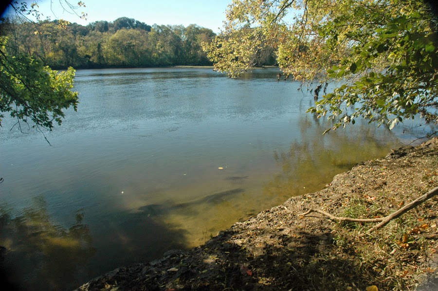
Last lab period I set up my Microaquarium. I obtained a glass rectangle glued on three sides with an opening at the top. This is where everything goes in. I chose to use water source #2, which was taken from the French Broad River located at the Seven Islands Wildlife Refuge.
 2. French Broad River, Seven Islands Wildlife Refuge, Kelly Lane , Knox Co. Tennessee. Partial shade exposure French Broad River Water Shed N35 56.742 W83 41.628 841 ft 10/12/2014
I took soil from the water source and added it to the bottom, and then took water from the bottom, middle, and top of the source. Two plants were then added to the Microaquarium. One was
-Amblestegium varium (Hedwig) Lindberg. Moss.
Collection from: Natural spring. at Carters Mill Park, Carter Mill Road, Knox Co. TN. Partial shade exposure. N36 01.168 W83 42.832. 10/12/2014,
and the other,
-Utricularia gibba L. Flowering plant. A
carnivorous plant. Original material from south shore of Spain Lake (N 35o55 12.35" W088o20' 47.00), Camp Bella Air Rd. East of Sparta Tn. in White Co. and grown in water tanks outside of greenhouse at Hesler Biology Building. The University of Tennessee. Knox Co. Knoxville TN.
10/12/2014
I then looked at the Microaquarium through a microscope and observed all of the living organisms. There were many including one giant worm (Nematode). Most moved very fast and spontaneously. It seemed as though the majority of life in my aquarium bunched up in the areas of the plants, which led me to believe they stay in the areas with the most oxygen. I am excited to see what develops in these aquariums.
French Broad River, Seven Islands Wildlife Refuge

I took soil from the water source and added it to the bottom, and then took water from the bottom, middle, and top of the source. Two plants were then added to the Microaquarium. One was
-Amblestegium varium (Hedwig) Lindberg. Moss.
Collection from: Natural spring. at Carters Mill Park, Carter Mill Road, Knox Co. TN. Partial shade exposure. N36 01.168 W83 42.832. 10/12/2014,
and the other,
-Utricularia gibba L. Flowering plant. A
carnivorous plant. Original material from south shore of Spain Lake (N 35o55 12.35" W088o20' 47.00), Camp Bella Air Rd. East of Sparta Tn. in White Co. and grown in water tanks outside of greenhouse at Hesler Biology Building. The University of Tennessee. Knox Co. Knoxville TN.
10/12/2014
I then looked at the Microaquarium through a microscope and observed all of the living organisms. There were many including one giant worm (Nematode). Most moved very fast and spontaneously. It seemed as though the majority of life in my aquarium bunched up in the areas of the plants, which led me to believe they stay in the areas with the most oxygen. I am excited to see what develops in these aquariums.
Subscribe to:
Comments (Atom)


